
St Emlyn’s Lesson Plans
Assessing acute coronary syndrome
Discuss urgent ECG findings, initial ACS care and the limits of high-sensitivity troponin pathways.
Early-career Emergency Medicine · 45-minute session
For learners
Before: Allow about 18 minutes in total.
When we meet: We will discuss an urgent ECG, a patient with a non-ischaemic ECG and how to explain the limits of reassuring tests.
For facilitators
Send: the learner preparation link before teaching.
Bring: Local chest-pain/ACS pathway, the ECG image set in Cases and discussion, a blank handover sheet and an optional approved full 12-lead ECG for case 2. A calculator is unnecessary unless using locally validated serial-result examples.
Before the session
If you are attending this session, complete the core preparation below before you come. Allow about 18 minutes in total.
Core preparation
NICE CG95: Recent-onset chest pain · About 8 minutes
Read CG95's acute-assessment recommendations on ECG and biochemical markers, including the limits of a normal initial ECG.
https://www.nice.org.uk/guidance/cg95/chapter/recommendations
NICE NG185: Acute coronary syndromes · About 7 minutes
Read NG185's initial-treatment and STEMI reperfusion recommendations. The facilitator will provide the local pathway for comparison during the case.
https://www.nice.org.uk/guidance/ng185/chapter/recommendations
Think about (allow 3 minutes): Which features would make you escalate before the first troponin result?
Picture the scene
A nurse brings you an ECG from a patient with chest pain. What do you need to know while you look at the tracing, and what must not wait for blood results?
By the end of the session
- Prioritise clinical assessment and ECG-based urgency while keeping alternative emergencies in view.
- Use the local ACS treatment and reperfusion pathway with patient-specific safety checks.
- Interpret hs-troponin as a marker within an assay-specific pathway, not as a universal test for every cardiac cause of pain.
After the session
- In your next chest-pain note, record which assay and pathway were used, what the results mean and any remaining clinical concerns.
Optional further learning
You do not need to complete this before attending. The listening time is additional to the core preparation above.
Optional listening · 19 min 30 sec
St Emlyn’s Podcast: Ep 299 - Which patients with chest pain need a troponin?
Explore how symptoms and clinical judgement influence the decision to request troponin.
Search for this episode by title in MedPod Learn if you want to record your listening there.
Identify one apparently reassuring feature that cannot safely exclude MI. Explain how a credible alternative diagnosis changes your assessment. The numerical estimates concern selected suspected-ACS populations; use the current local ECG/troponin pathway. If you have heard this episode before, bring your previous reflection.
https://www.stemlynspodcast.org/e/ep-299-which-patients-with-chest-pain-need-a-troponin/
Facilitator guide
Send learners the preparation link several days before teaching. Allow about 18 minutes in total.
Before learners arrive
- Confirm the local assay, reporting units, sampling times and pathway. Check urgent cardiology/reperfusion contacts and the locally approved initial treatment chart.
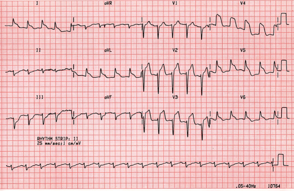
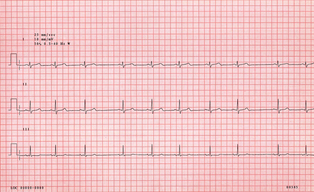
- Enlarge ECG 1 for the urgent case. ECG 2 contains only leads I–III: use it to discuss rhythm and the limits of incomplete recordings. Supply an approved complete 12-lead ECG if visual practice is wanted for case 2; otherwise use the explicit text assumption.
Equipment: Local chest-pain/ACS pathway, the ECG image set in Cases and discussion, a blank handover sheet and an optional approved full 12-lead ECG for case 2. A calculator is unnecessary unless using locally validated serial-result examples.
Setup: Work in threes: referring clinician, cardiology registrar and observer. Each learner rehearses the first 30 seconds of the urgent referral; the observer checks urgency, uncertainty and the requested action. For the second case, take turns explaining the assessment to the patient.
If the preparation has not been completed
Replace the five-minute opening with the two written case summaries and the local chest-pain/reperfusion pathway. Ask pairs which information could change action immediately and which can be gathered in parallel. Keep the pathway open before discussing medicines. In the 30-minute plan, use five minutes here and reduce the urgent case from 10 to eight minutes.
Suggested session plan
- 0–5 min — The ECG at the desk. Ask what needs to happen now and which questions can run alongside it.
- 5–19 min — Time-critical case. Rehearse the call and patient-specific treatment checks without waiting for a biomarker.
- 19–34 min — Normal ECG and troponin. Explore exactly what the pathway rules out and why persistent concern matters.
- 34–40 min — Patient explanation. Practise a discharge explanation with a clear safety net and no zero-risk promise.
- 40–45 min — Close. Ask the group to replace the phrase “troponin negative, discharge” with an informative clinical summary.
Start the discussion
Read the opening scenario together. Give everyone a moment to think, then ask what concerns them and what they need to know next.
Cases and discussion
Use these cases, or invite an anonymised case from the group that addresses the same questions.
Teaching image set
Useful during the session: teaching images
Ongoing pain and an ischaemic ECG
A 50-year-old has ongoing central chest pain radiating to the arm and throat and looks clammy. Review ECG 1 before opening the discussion notes.
Add this information as the discussion develops:
- Ask learners for the next three actions without offering a troponin result.
- The blood pressure is lower than expected and the patient asks for pain relief.
- A colleague reports the pain was abrupt and maximal at onset; ask what competing diagnoses need consideration while help is mobilised.
Ask the group:
- Which findings make this time critical?
- What can happen in parallel with the reperfusion call?
- Which patient-specific checks matter before antiplatelets, nitrates or opioid analgesia?
- How would a possible aortic presentation alter the conversation with the senior and cardiology team?
Discussion notes
Points to draw out:
- ECG 1 shows anterior ST elevation with reciprocal inferior change. The tracing alone cannot establish whether these changes are new; interpret it with the history and any previous ECG.
- Obtain urgent senior/cardiology input and use the reperfusion pathway when STEMI is suspected; do not wait for a troponin result to begin time-critical assessment and escalation.
- NICE NG185 supports prompt aspirin for ACS unless contraindicated, with further antithrombotic and reperfusion decisions following the appropriate pathway. Analgesia and haemodynamic assessment remain important; treatment is not a fixed mnemonic detached from blood pressure and contraindications.
- The ECG must be interpreted with the presentation. A competing emergency such as aortic dissection can change the risk of standard treatment and needs immediate senior discussion.
Common misconceptions:
- Waiting for biomarkers despite a time-critical ECG.
- Treating an ECG in isolation from the person and history.
For a more experienced group: Ask a senior group to coordinate a transfer when the usual PCI destination is temporarily unavailable.
Normal ECG, ongoing uncertainty
A 35-year-old has intermittent chest discomfort, smokes occasionally and wants reassurance that their age makes a heart attack impossible. A complete 12-lead ECG has been assessed as non-ischaemic. ECG 2 is a separate three-lead rhythm illustration; it cannot establish that finding.
Add this information as the discussion develops:
- Ask for a focused history, examination and alternative diagnoses before choosing a test.
- The first hs-troponin is below the laboratory’s stated threshold, but the sample was taken early and the local pathway may require another sample.
- In a variation, troponin is raised in a patient with renal impairment and a concurrent acute illness.
Ask the group:
- What can the three visible leads in ECG 2 demonstrate, and what further recording is needed to assess possible ischaemia?
- What does youth change, and what does it not establish?
- What exact pathway, assay and timing are being used?
- Does a low result rule out every ACS presentation or every cardiac cause of pain?
- What information distinguishes myocardial injury from an acute coronary mechanism?
- What should the patient understand before leaving?
Discussion notes
Points to draw out:
- A normal ECG does not exclude ACS. Use the local validated hs-troponin pathway, including sampling intervals, thresholds and clinical assessment. Some selected pathways allow a single very low result, but “negative troponin” is not a universal discharge rule.
- Raised troponin indicates myocardial injury in the appropriate clinical context; it is not synonymous with type 1 MI. Serial change, symptoms, ECG and the clinical setting inform interpretation. Renal impairment does not make a result meaningless or automatically explain it away.
- Explain what has been assessed, what remains uncertain, the likely alternative explanation where possible, and specific return/follow-up advice.
Common misconceptions:
- Calling a troponin rule-out “no cardiac cause”.
- Dismissing a raised result solely because kidney function is abnormal.
- Using absence of conventional risk factors to close the assessment.
For a more experienced group: Use a pair of fictional serial results from the locally approved teaching pathway and ask what additional information is still needed.
Important clinical points
- Do not delay urgent reperfusion assessment for troponin.
- Do not generalise an assay-specific rule-out threshold across laboratories.
- Consider serious alternative causes of chest pain before routine ACS treatment or discharge.
- A regular sinus rhythm strip or three-lead extract cannot exclude ischaemia; obtain and interpret a full 12-lead ECG in suspected ACS.
Closing the session
Use these questions to check what the group will take away. Go back over any clinical points that remain unclear.
- When should a blood result not be awaited?
- What does a raised troponin prove and not prove?
- How would you explain the limits of a reassuring pathway result?
Key teaching points:
- Act on a time-critical presentation without waiting for troponin.
- Interpret troponin using the correct assay, sampling times and clinical context.
- Before discharge, consider remaining clinical concerns and explain what the assessment has and has not excluded.
Before leaving, ask each person to choose one action from After the session and say when they will try it. They complete that follow-up in practice, with supervision where needed.
Adapting the discussion
Uncertainty and local practice
Symptoms overlap, assays differ and some patients need further assessment despite initially reassuring tests. Use the numerical thresholds and sampling times in the approved local pathway.
If the discussion drifts
What decision are you making now, and what information actually supports it?
For a more experienced group
Discuss myocardial injury, type 2 MI and type 1 MI using a locally reviewed case, including the risk of both overtreatment and premature dismissal.
A 30-minute version
30-minute option: opening 3, urgent case 10, troponin case 12, explanation and closing 5. Omit transfer logistics.
Evidence and guidance
- NICE CG95: Recent-onset chest pain — Assessment, ECG and hs-troponin in clinical context; a normal ECG does not exclude ACS.https://www.nice.org.uk/guidance/cg95/chapter/recommendations
- NICE NG185: Acute coronary syndromes — Current ACS treatment pathway, including reperfusion and antithrombotic decisions.https://www.nice.org.uk/guidance/ng185/chapter/recommendations
- NICE HTG552: High-sensitivity troponin tests — Assay-specific guidance for early NSTEMI rule-out, previously published as DG40.https://www.nice.org.uk/guidance/htg552
Suggested curriculum links
RCEM 2021 curriculum (2025 update): SLO1 Adult acute assessment
The cases integrate symptoms, ECG and biomarker interpretation.
RCEM 2021 curriculum (2025 update): SLO3 Adult resuscitation
The urgent case practises escalation and stabilisation for suspected time-critical ACS.
Check these links against the learner’s stage and programme. Attendance at a teaching session does not establish competence.
First published 2020-05-11 · Iain Beardsell.